
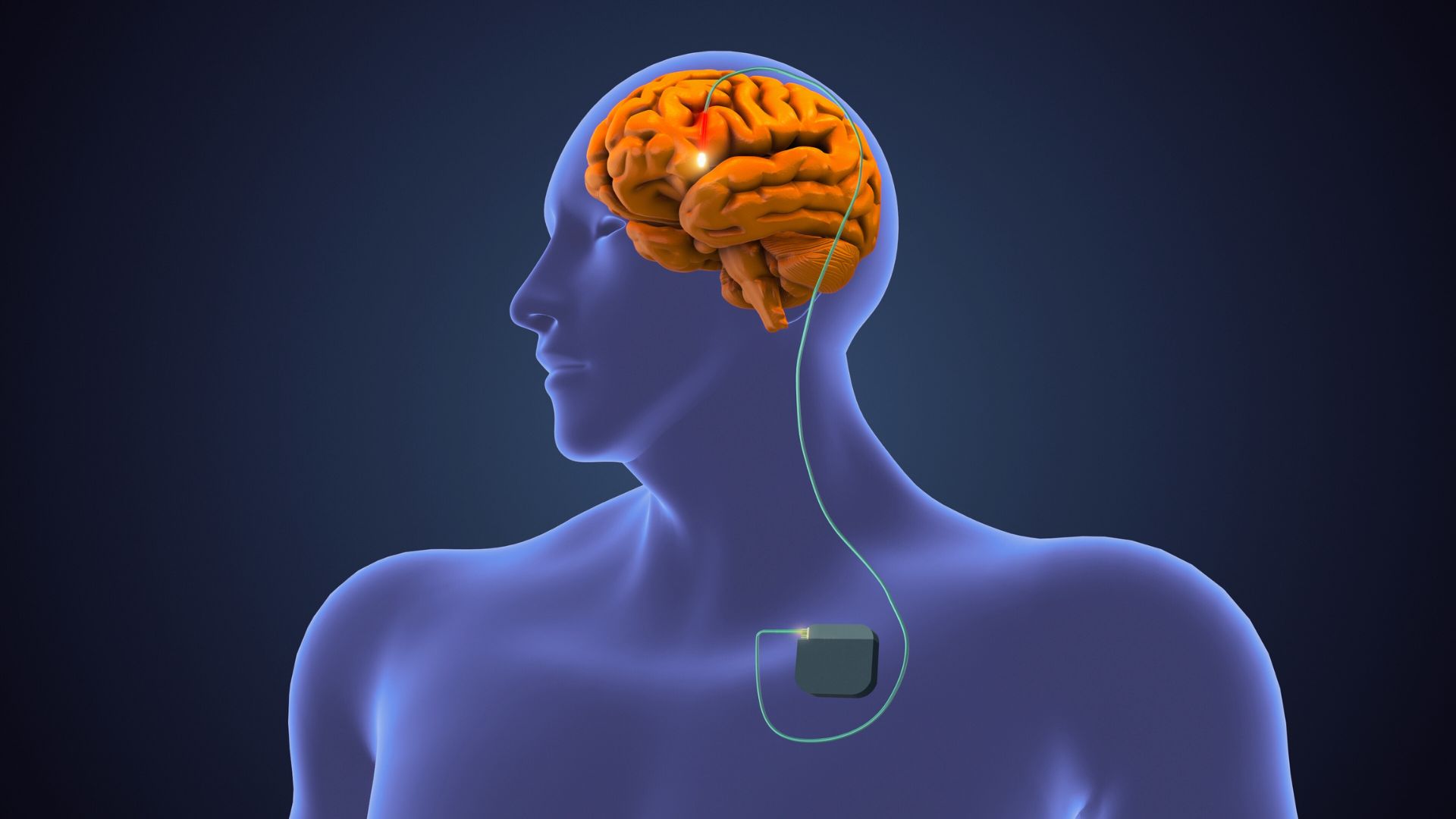
The effects of brain injury are largely dependent on the location and severity of the trauma. The brain is a complex organ composed of three main parts; the cerebrum, the cerebellum and the brainstem. These areas communicate via billions of neurons to control many of the body’s functions, like movement, speech, thoughts, and emotions.
In this article, we look at the structure and function of these parts of the brain and the symptoms that may result due to injury.
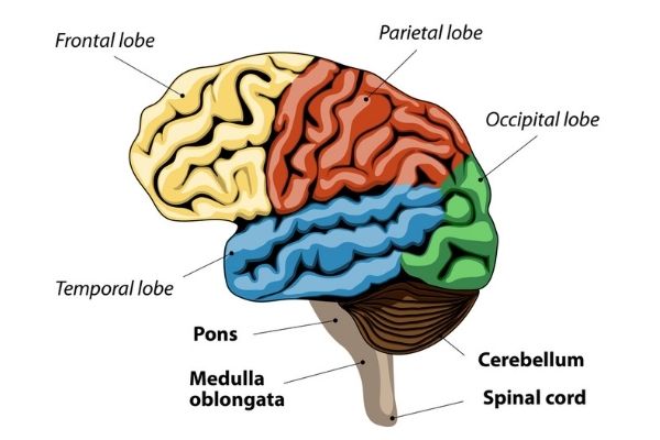
Cerebrum Anatomy and Function
The largest part of the human brain is the cerebrum which is divided into right and left sides (hemispheres). These hemispheres are joined by a bundle of nerve fibers called the corpus collosum. The right and left hemispheres of the brain communicate via these nerve fibers. Each hemisphere controls the opposite side of the body. When a stroke occurs in one side of the brain the opposite side of the body is affected.
Each hemisphere contains four different lobes each with unique functions:
Frontal Lobe Injury
The frontal lobe controls executive functioning such as planning, problem solving, memory, language, attention and voluntary movements. Our frontal lobes are responsible for many of our behaviours and is part of our emotional control centre.
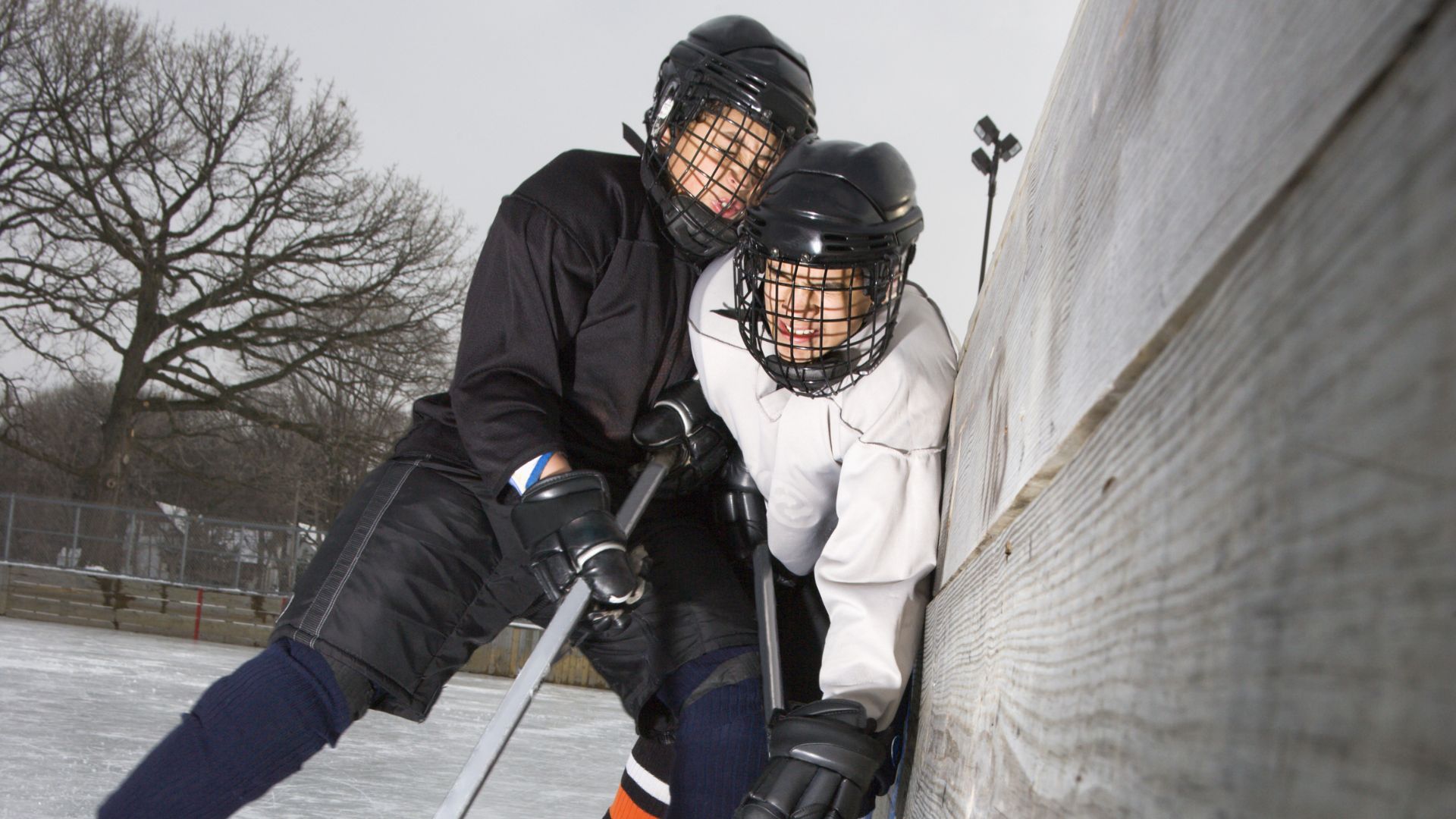
Injury to the frontal lobe through a traumatic brain injury or stroke can cause a range of difficulties depending on the location and extent of the injury. This includes paralysis, speech difficulties such as aphasia, impaired judgement, difficulty problem solving, mood changes, decreased motivation and impulsive behaviour.
The frontal lobe is fully developed at 25 years of age.[i] Due to its location and size the frontal lobe is particularly vulnerable to head trauma.
Parietal Lobe Injury
The parietal lobe is responsible for processing information from the body including touch, temperature, pain and joint position. It is responsible for integrating this sensory information to provide a picture of the world around us.
Injury to this area of the brain can also have a wide range of effects including lack of awareness of certain body parts/space (neglect), difficulty with motor planning (apraxia), reading and writing deficits as well as an inability to process sensations such as hot and cold.[ii]
Temporal Lobe Injury
This temporal lobe is associated with processing auditory information and the encoding of memory.
Injury to the temporal lobe can result in difficulties understanding spoken words (receptive aphasia), impaired selective attention and difficulty learning and retaining new information.[iii]
Occipital Lobe Injury
The occipital lobe is the visual processing area of the brain. It is associated with visuospatial processing, depth perception, colour distinction, object and face recognition and memory formation.[iv]
Injury to this part of the brain can result in visual inattention, blindness or blind spots, and hallucinations.
Cerebellum Anatomy, Function & Injury
The cerebellum is located at the back of the brain under the occipital and temporal lobes. Although the cerebellum accounts for only 10 percent of the brain’s volume, it contains over 50 percent of the total number of neurons in the brain.[v] The cerebellum has many roles including:
- Maintenance of balance and posture
- Coordination of voluntary movement
- Motor learning
- Cognitive functions[vi]
With injury to the cerebellum movement and balance are typically affected. Cerebellar ataxia can occur due to many diseases and is associated with difficulties in walking, balance, coordination of movement of the limbs and difficulty with eye movements.
Brainstem Anatomy, Function & Injury
The brainstem is at the bottom of the brain and connects the brain to the spinal cord. It consists of three parts: midbrain, pons and medulla oblongata. The brainstem helps to coordinate messages that relate to:
- Heart rhythms
- Blood pressure
- Balance
- Breathing
- Swallowing
- Facial sensations
- Balance[vii]
Injury to the brainstem can range from symptoms of nausea, inability to cough, dizziness, and abnormal sleep patterns. Serious injury to the brainstem can result in coma and death due to its role in vital bodily functions.
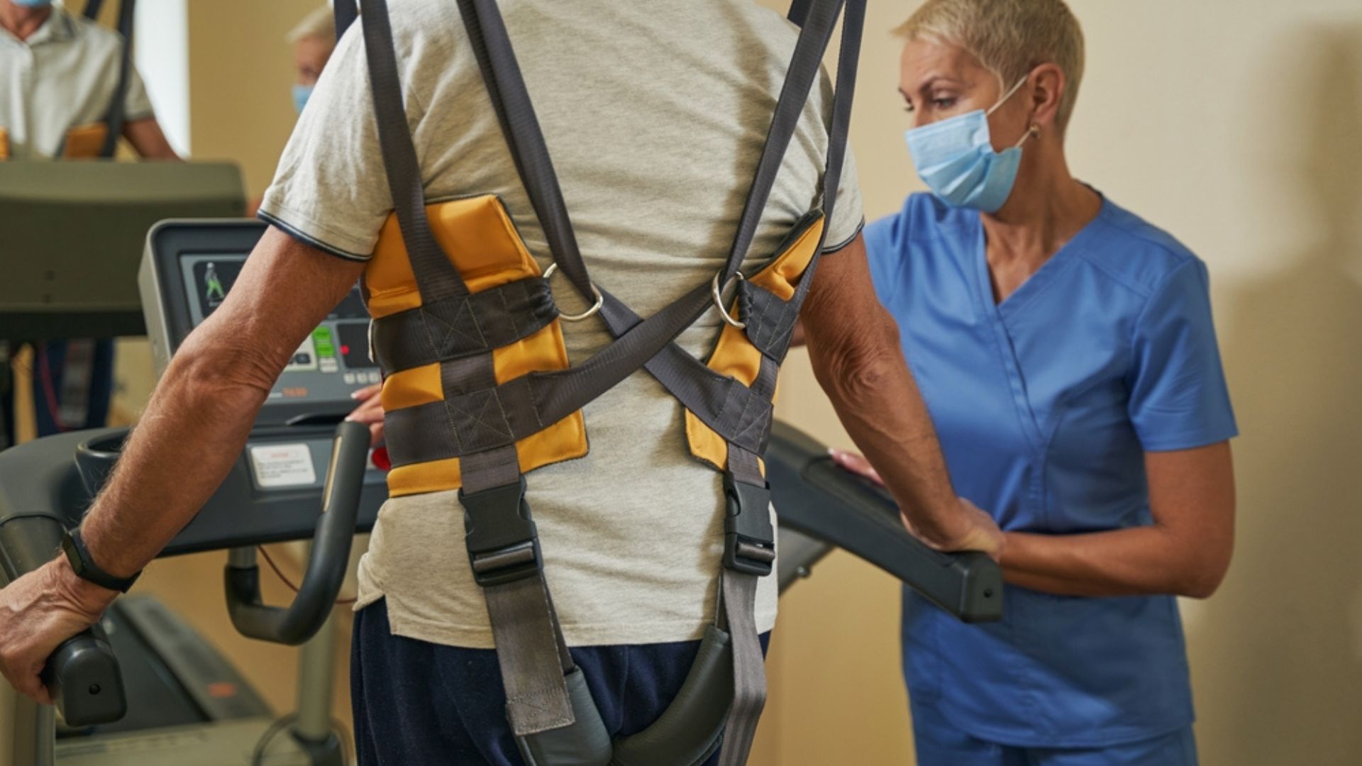
Neuroplasticity and Neurological Injury Rehabilitation
Although each area of the brain is often discussed separately, it is important to note there is tremendous overlap in the function of these structures. It is also important to remember that the architecture of the brain is made up of billions of neurons that communicate between the different areas of the brain. We now know that the brain has an amazing ability to adapt, modify, and change both structure and function to experiences.
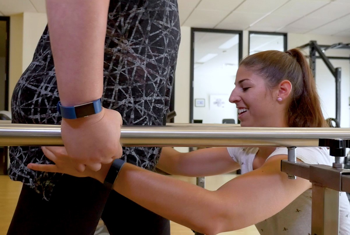
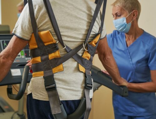
At Propel Physiotherapy, we use the principles of neuroplasticity and motor learning to treat the effects of brain injuries. Through proper therapy and training the brain has tremendous ability to change and repair. This can translate to improved function and independence for the client.
References
[i] Arain M, Haque M, Johal L, et al. Maturation of the adolescent brain. Neuropsychiatr Dis Treat. 2013;9:449-461. doi:10.2147/NDT.S39776
[ii] Jeffrey M. Clarke, Neuroanatomy, 3 The Parietal Lobes, in Neuropsychology, 1994
[iii] Brain Map: Temporal Lobes, Queensland Health, Queensland Government
[iv] Rehman A, Al Khalili Y. Neuroanatomy, Occipital Lobe. [Updated 2021 Jul 31]. In: StatPearls [Internet]. Treasure Island (FL): StatPearls Publishing; 2022 Jan-. Available from: https://www.ncbi.nlm.nih.gov/books/NBK544320/
[v] James Knierim, Ph.D., Overview: Functions of the Cerebellum, Department of Neuroscience, The Johns Hopkins University
[vi] James Knierim, Ph.D., Overview: Functions of the Cerebellum, Department of Neuroscience, The Johns Hopkins University
Written by


